In Vitro vs In Vivo: What’s the Difference in Research Studies?

By Dr. Leonard Haberman, Chief Science Officer, OPTMZ Peptides | Published: April 2, 2026 | Last updated: April 17, 2026 In vitro research is performed outside a living organism — in controlled environments like cell cultures, isolated tissue preparations, or purified molecular systems. In vivo research is performed inside a living organism, such as an animal model. In vitro studies isolate a single variable under highly controlled conditions; in vivo studies observe how a compound behaves within a complete biological system. Both methods serve distinct scientific purposes, and rigorous preclinical peptide research typically progresses from one to the other rather than choosing between them. The terms themselves come from Latin: in vitro translates roughly to “in glass,” reflecting the historical use of test tubes and petri dishes, while in vivo translates to “in the living.” In modern research, the distinction is methodological rather than literal — in vitro systems now include sophisticated organ-on-chip platforms and 3D organoids, while in vivo encompasses everything from zebrafish models to non-human primate studies. What Does In Vitro Mean in a Research Context? An in vitro experiment takes place in a controlled artificial environment outside a living organism. Researchers use in vitro systems to study specific biological interactions in isolation — free from the confounding variables present in a whole organism. The goal is mechanistic precision. Common in vitro systems in peptide research include: Cell culture — immortalized cell lines (e.g., HEK293, HepG2, C2C12) or primary cell preparations maintained in growth media Receptor binding assays — isolated or membrane-bound receptor preparations used to characterize peptide-receptor affinity and selectivity Enzyme kinetics assays — purified enzyme systems used to measure substrate turnover or inhibition 3D organoid and spheroid cultures — multi-cellular constructs that more closely mimic tissue-level behavior Organ-on-chip platforms — microfluidic devices that recreate organ-level physiology in vitro In peptide research, in vitro assays are often the first experimental step after a compound has been synthesized and characterized. For example, studies investigating the tripeptide GHK-Cu have used in vitro fibroblast cultures to examine its interactions with collagen gene expression pathways (Pickart & Margolina, 2018 — PubMed). What Does In Vivo Mean in a Research Context? In vivo research is conducted inside a living organism. Rather than isolating one variable, in vivo studies observe how a compound interacts with the full biological context of a functioning system — circulation, metabolism, organ crosstalk, immune response, and tissue distribution. Common in vivo model systems include: Rodent models — mice and rats, used for the majority of preclinical pharmacology research Zebrafish — increasingly used for early-stage developmental and cardiovascular studies Non-human primates — reserved for late-stage preclinical work where physiological similarity to humans is essential Ex vivo preparations — tissues removed from a living organism and studied shortly after, occupying a middle category between in vitro and in vivo Peptide research frequently progresses from in vitro characterization into in vivo rodent work. BPC-157, a synthetic pentadecapeptide derived from a protective protein found in gastric juice, has been studied in rodent models for its effects on connective tissue healing kinetics (Chang et al., 2011 — PubMed; Seiwerth et al., 2018 — PubMed). These rodent studies followed earlier in vitro work characterizing the peptide’s interactions at the cellular level. How Do In Vitro and In Vivo Studies Fit in the Preclinical Research Progression? In modern peptide research, in vitro and in vivo approaches are rarely an either/or choice. They occupy different stages of a staged research progression: In silico — computational modeling of peptide structure, receptor docking, and predicted binding affinities. This stage requires no physical compound and is used to prioritize candidates for synthesis. In vitro — characterized peptide is tested in purified or cell-based systems to establish mechanism, selectivity, and dose-response behavior in isolation. Ex vivo — excised tissue or organ preparations bridge the gap between cell-based in vitro work and whole-organism in vivo work. In vivo — compound is evaluated in living animal models to assess pharmacokinetics, tissue distribution, and system-level effects. Clinical research — where applicable, and only after extensive preclinical work, compounds advance into human clinical trials under regulatory oversight. Each stage answers questions the previous stage could not. In vitro assays can demonstrate that a peptide binds its intended receptor with nanomolar affinity — but only in vivo work can show how that binding translates into distribution, metabolism, and physiological outcomes in a whole organism. In vivo work, in turn, cannot substitute for the mechanistic clarity that in vitro work provides. What Are the Strengths and Limitations of Each Approach? Each methodology offers distinct advantages and carries distinct constraints. The table below summarizes how in vitro and in vivo approaches compare across the dimensions researchers weigh when designing a preclinical study. Dimension In Vitro In Vivo What it measures Isolated mechanism under controlled conditions Compound behavior in a complete biological system Variable control High — single receptor, cell type, or pathway studied in isolation Low — biological complexity introduces many confounding factors Throughput High — large compound libraries screened in parallel Low — labor-intensive per subject Cost per data point Lower Higher Reproducibility High — controlled conditions yield consistent results Lower — biological variability between subjects Biological complexity Reduced — lacks tissue architecture, vascular supply, cross-system signaling Full — captures immune, endocrine, and metabolic integration Pharmacokinetics (ADME) Not captured Captured — absorption, distribution, metabolism, excretion Translational accuracy Indirect — in vitro potency often does not correspond to in vivo activity Closer to physiological reality; species differences remain Animal use None for primary screening Required, with ethical oversight Typical stage of use Early mechanistic characterization and screening Later-stage validation and system-level evaluation The methodological tradeoff: in vitro studies tell you what a compound can do; in vivo studies tell you what it does under real biological conditions. How Is In Vitro and In Vivo Research Applied to Peptide Compounds? Peptide compounds present particular methodological considerations across the in vitro / in vivo continuum. Stability and half-life. Many peptides degrade rapidly under physiological conditions. In vitro
Acetic Acid 0.6% for Research Peptides: When to Use It, How It Works, and How OPTMZ Verifies the Solvent

Acetic acid is a widely used compound in chemical and laboratory settings, playing a role in everything from synthesis processes to analytical applications. As research continues to evolve, new methods are improving how acetic acid is produced, making the process more efficient, scalable, and environmentally conscious. What Is Acetic Acid? Acetic acid is a simple organic compound known for its role in various chemical reactions. In research and industrial settings, it’s commonly used as a solvent, reagent, and intermediate in synthesis. Its versatility makes it an important component in both small-scale laboratory work and large-scale production systems. Traditional Production Methods Historically, acetic acid has been produced through processes such as: Methanol carbonylation Oxidation of acetaldehyde Fermentation-based methods Among these, methanol carbonylation has become the dominant industrial approach due to its efficiency and scalability. What’s Changing in 2025 Recent developments are focused on improving sustainability, reducing waste, and increasing precision in production. Advanced CatalystsNew catalyst systems are being developed to improve reaction efficiency and reduce energy consumption. These catalysts help speed up production while minimizing unwanted byproducts. Greener Production TechniquesThere is a growing shift toward environmentally friendly methods, including bio-based production and reduced reliance on harsh chemicals. Process OptimizationModern systems are using improved monitoring and control technologies to ensure more consistent output and better resource management. Integration with Renewable InputsSome research is exploring the use of renewable feedstocks, aiming to reduce dependence on traditional petrochemical sources. Why These Innovations Matter Improving how acetic acid is produced has a direct impact on research and manufacturing. Better processes mean: Higher purity and consistency Reduced environmental impact Lower production costs over time More reliable supply chains For laboratories and industries that rely on precise chemical inputs, these improvements make a noticeable difference. Applications in Research Acetic acid continues to be used across various research areas, including: Chemical synthesis Analytical testing Peptide and organic compound preparation Buffer and solvent systems Its role as a foundational compound ensures it remains relevant as research techniques advance. Final Thoughts Acetic acid synthesis is evolving with a clear focus on efficiency and sustainability. Innovations in catalysts, production methods, and process control are shaping how this essential compound is produced and used. As research continues, these advancements are expected to further improve both quality and environmental impact, making acetic acid production more refined and future-ready.
Peptide Mass Spectrometry: How Identity Verification Works in Research-Grade Compound Supply
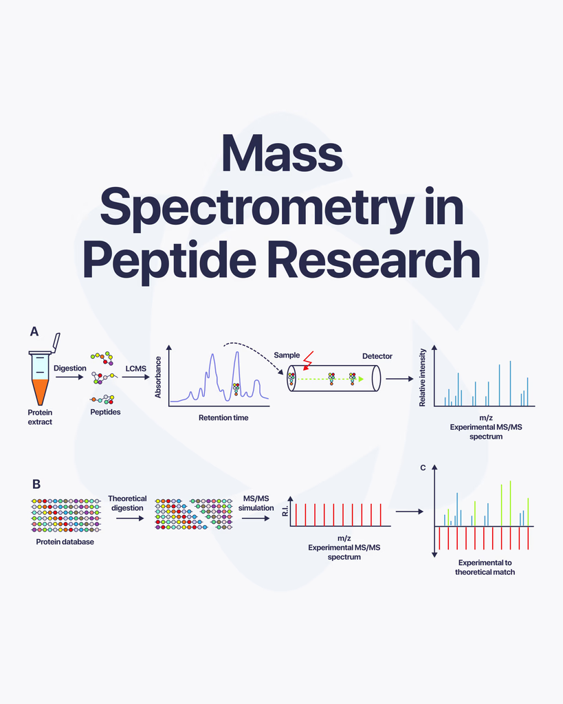
By Dr. Leonard Haberman, Chief Science Officer, OPTMZ Peptides Published: March 2,2026 | Last Updated: April 13, 2026 Peptide mass spectrometry is an analytical technique that determines the molecular identity of a peptide by measuring the mass-to-charge ratio of its ionized fragments. In research-grade compound supply, it is the definitive method for confirming that a peptide’s amino acid sequence matches its specified structure — a function that HPLC purity analysis alone cannot perform. What Is Peptide Mass Spectrometry? Mass spectrometry (MS) measures the mass-to-charge ratio (m/z) of ionized molecules. When applied to peptides, this produces a spectrum of mass values that functions as a molecular fingerprint: each peptide generates a unique pattern of fragment ions that corresponds to its specific amino acid sequence and molecular weight. In the context of research-grade peptide verification, mass spectrometry answers a question that purity data cannot: is this compound actually the peptide it is labeled as? A sample can achieve 99% purity by HPLC and still be the wrong compound — or a structurally similar analog — if identity confirmation is not independently performed. Mass spectrometry closes that verification gap. The technique was first systematically applied to peptide fragmentation analysis by Roepstorff and Fohlman, whose 1984 nomenclature for peptide fragment ion series (b-ions and y-ions) established the interpretive framework still in use today (Roepstorff P & Fohlman J, 1984, PMID: 6525415). How Does Mass Spectrometry Identify a Peptide? The identification process proceeds in three stages: 1. Ionization The peptide sample must be converted to gas-phase ions before mass measurement. The two dominant ionization methods for peptides — ESI and MALDI — are described in detail below. Ionization method selection affects sensitivity, throughput, and the types of structural information extractable from the spectrum. 2. Mass separation Ions are separated in a mass analyzer based on their m/z ratio. Common analyzer types in peptide MS include quadrupole, time-of-flight (TOF), and ion trap instruments. Each offers different trade-offs between mass accuracy, resolution, and scan speed. For high-resolution research-grade identity confirmation, TOF and Orbitrap analyzers are typically used due to their sub-ppm mass accuracy. 3. Detection and spectrum interpretation The detector produces a spectrum of ion abundances across m/z values. For intact peptide identity confirmation, the molecular ion peak is matched against the theoretical mass of the target compound. For sequence-level confirmation via tandem MS, the fragment ion series is mapped against predicted b-ion and y-ion series for the stated amino acid sequence. A match between observed and theoretical mass within the instrument’s accepted error tolerance (typically ≤5 ppm for high-resolution instruments) constitutes identity confirmation. Deviation beyond this tolerance indicates a structural mismatch, degradation product, or synthesis error — all of which constitute grounds for batch rejection in a quality-controlled supply chain. What Is the Difference Between ESI and MALDI for Peptide Analysis? The two primary ionization methods used in peptide mass spectrometry each have distinct operational characteristics and applications: Electrospray Ionization (ESI) ESI converts peptides in solution directly to gas-phase ions by passing the liquid sample through a charged capillary under atmospheric pressure. The process generates multiply charged ions, which is particularly useful for larger peptides — the multiple charge states allow heavier molecules to appear at lower m/z values within the instrument’s detection range. ESI is highly compatible with liquid chromatography (LC), making LC-MS and LC-MS/MS the dominant platform for comprehensive peptide analysis. The continuous-flow nature of ESI allows real-time coupling with separation systems, enabling simultaneous purity profiling and identity confirmation in a single analytical run. Yates, Ruse, and Nakorchevsky (2009) provide a comprehensive overview of LC-MS/MS workflows in proteomics and research compound characterization (PMID: 19838170). At Krause Analytical, ESI-based mass spectrometry is used as part of the identity confirmation step in OPTMZ Peptides’ testing panel — conducted on every batch submitted for quality verification, independently of purity analysis. Matrix-Assisted Laser Desorption/Ionization (MALDI) MALDI embeds the peptide sample in a crystalline matrix material that absorbs laser energy. When the laser fires, the matrix transfers that energy to the peptide molecules, desorbing and ionizing them from the solid surface. MALDI typically produces singly charged ions and is particularly well-suited to rapid molecular weight determination and peptide mass fingerprinting (PMF). MALDI instruments — especially MALDI-TOF platforms — are widely used in high-throughput screening environments due to their speed and relative tolerance for sample contaminants. However, for sequence-level confirmation requiring tandem fragmentation, ESI-based systems are generally preferred in research-grade supply chain applications due to their direct compatibility with MS/MS workflows. What Does MS/MS (Tandem Mass Spectrometry) Add to Peptide Sequencing? Standard MS provides a molecular weight measurement — confirmation that the peptide falls within the expected mass range. Tandem mass spectrometry (MS/MS) goes further: it fragments the parent ion and analyzes the resulting fragment ions to reconstruct the amino acid sequence directly. The MS/MS process works as follows: Precursor ion selection: A specific m/z value (the molecular ion of interest) is isolated in the first mass analyzer stage Fragmentation: The isolated ion is subjected to collision-induced dissociation (CID), electron transfer dissociation (ETD), or higher-energy collisional dissociation (HCD), breaking the peptide backbone at specific sites Fragment ion analysis: The resulting fragment ions are analyzed in the second stage, generating a spectrum of b-ions (N-terminal fragments) and y-ions (C-terminal fragments) Sequence reconstruction: The mass differences between adjacent b-ions or y-ions correspond to the masses of individual amino acid residues, allowing direct sequence readout Syka et al. (2004) demonstrated the utility of electron transfer dissociation for sequence analysis of peptides and proteins, establishing an important methodological extension to conventional CID-based MS/MS (PMID: 15258601). For research-grade peptide suppliers, MS/MS is the gold standard for verifying that a compound’s primary structure — its actual amino acid sequence — matches the label claim, not merely its molecular weight. How Is Mass Spectrometry Used in Research-Grade Peptide Quality Control? In a controlled peptide supply chain, mass spectrometry performs two non-redundant functions that together constitute complete identity verification: Function 1: Intact mass confirmation The intact molecular ion mass is compared to the