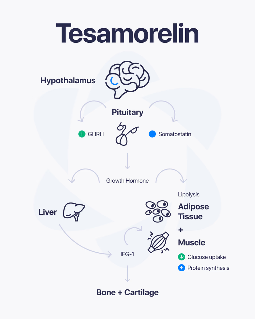
By Dr. Leonard Haberman, Chief Science Officer, OPTMZ Peptides Published April 18, 2026 · Last updated April 18, 2026 Tesamorelin is a synthetic 44-amino-acid analog of human growth hormone-releasing hormone (GHRH), originally developed under the code TH9507. It is chemically designated N-(trans-3-hexenoyl)-[Tyr¹]hGRF(1-44)NH₂ acetate — a GHRH(1-44) sequence modified at the N-terminal tyrosine with a hexenoyl fatty acid group that resists proteolytic cleavage. This modification is the central reason tesamorelin has been studied as a stabilized pharmacological probe for GHRH receptor signaling in research contexts (Ferdinandi et al., 2007, PMID 17214611). This page summarizes the molecular structure, receptor pharmacology, published research literature, and analytical verification standards relevant to tesamorelin as a research-grade peptide. All references are cited from peer-reviewed sources. Mechanistic claims are anchored to PubMed-indexed studies. What Is Tesamorelin? Tesamorelin is a synthetic growth hormone-releasing factor (GRF) analog derived from the full 44-amino-acid sequence of human GHRH (hGRF). Unlike shorter GHRH-pathway peptides such as sermorelin (GHRH 1-29), tesamorelin retains the complete native sequence and adds a structural modification at the N-terminus that prevents rapid enzymatic degradation. The compound activates the GHRH receptor (GHRH-R), a G protein-coupled receptor (GPCR) expressed on pituitary somatotropes. Receptor binding drives pulsatile release of endogenous growth hormone (GH) rather than introducing exogenous GH directly — a pharmacological distinction that has made it an object of research interest in studies of the GH/IGF-1 axis (Wang, 2009, PMID 19243281). The Molecular Structure of Tesamorelin Tesamorelin’s full chemical designation is N-(trans-3-hexenoyl)-[Tyr¹]hGRF(1-44)NH₂ acetate. Breaking this down: hGRF(1-44)NH₂ — the 44-amino-acid sequence of human growth hormone-releasing factor, C-terminally amidated [Tyr¹] — tyrosine at position 1, retained from native GHRH N-(trans-3-hexenoyl) — a six-carbon unsaturated fatty acid (hexenoyl) group covalently attached to the N-terminus acetate — the salt form used in pharmaceutical and research-grade preparations The trans-3-hexenoyl modification is the functional innovation. Native GHRH is cleaved rapidly by the enzyme dipeptidyl peptidase-IV (DPP-IV) at the Tyr¹-Ala² bond, producing the inactive GHRH(3-44) metabolite within minutes of administration. Attaching the hexenoyl group to the N-terminal tyrosine sterically blocks DPP-IV access, extending the molecule’s functional half-life in plasma (Ferdinandi et al., 2007). The molecular weight of tesamorelin acetate is approximately 5,196 Da, which places it in the mid-range for synthetic peptide research compounds and informs HPLC method selection for purity verification. How Does Tesamorelin Interact With the GHRH Receptor? Tesamorelin binds the GHRH receptor (GHRH-R), a class B GPCR located primarily on somatotroph cells in the anterior pituitary. The signal cascade has been characterized as follows in published research: Receptor binding: Tesamorelin engages the extracellular N-terminal domain and transmembrane core of GHRH-R, mimicking the binding pose of native GHRH. Gαs activation: Conformational change activates the heterotrimeric Gs protein, stimulating adenylate cyclase. cAMP elevation: Intracellular cAMP rises, activating protein kinase A (PKA) and CREB-mediated transcription of the GH gene. Pulsatile GH release: Somatotropes release stored GH into circulation in discrete pulses — a pattern that preserves the native pulsatility of endogenous GH secretion rather than producing sustained hormone elevation. Hepatic IGF-1 induction: Circulating GH binds hepatic GH receptors, driving synthesis and release of insulin-like growth factor-1 (IGF-1), the major downstream effector of the GH axis. Stanley and colleagues observed that tesamorelin administration in a controlled research population preserved physiological pulsatile GH secretion patterns and did not produce the insulin resistance commonly reported with exogenous GH administration (Stanley et al., 2011, PMID 20943777). This finding is frequently cited as the mechanistic rationale for classifying tesamorelin as a GHRH secretagogue rather than a GH replacement analog. What Has Research Examined About Tesamorelin? Published tesamorelin research spans pharmacokinetic characterization, GH-axis modulation, and metabolic parameter analysis. The following areas are well-represented in the peer-reviewed literature: GH and IGF-1 Axis Modulation Multiple controlled studies have measured circulating GH and IGF-1 changes following tesamorelin administration. Stanley et al. (2011) documented preserved pulsatile GH secretion and a rise in mean IGF-1 levels without evidence of the supraphysiological IGF-1 surges associated with recombinant GH protocols. Pharmacokinetics and Metabolic Parameters Clemmons and colleagues reported 12-month data on tesamorelin pharmacokinetics and metabolic parameters (including glucose tolerance markers) in a controlled research population (Clemmons et al., 2017, PMC5472315). The dataset remains one of the longest continuous research observations of GHRH-pathway modulation published in the analog class. Clinical Literature on HIV-Associated Lipodystrophy The majority of published tesamorelin human research has been conducted in the context of HIV-associated lipodystrophy, a condition characterized by altered regional adipose distribution. Spooner and Olin (2012, PMID 22298602) reviewed the clinical trial literature in this population, and a 2024 meta-analysis examined randomized controlled trial outcomes across the literature base (Badran et al., 2024). OPTMZ Peptides reports this research record as documentary reference — we do not promote tesamorelin as a consumer product for any indication. Broader Metabolic Research Recent review literature has situated tesamorelin within the larger context of GHRH-pathway research in diabetes and metabolic physiology (Steenblock & Bornstein, 2024, PMC12137473). Ongoing research continues to characterize GHRH receptor distribution in peripheral tissues and the role of local GHRH signaling outside the pituitary. Tesamorelin vs. Native GHRH: Pharmacokinetic Differences Parameter Native GHRH Tesamorelin Amino acid length 44 (hGRF 1-44) 44 (hGRF 1-44) N-terminal modification None trans-3-hexenoyl group DPP-IV resistance No — cleaved at Tyr¹-Ala² Yes — hexenoyl blocks access Plasma half-life ~6-7 minutes ~26-38 minutes (published range) Receptor affinity Reference Comparable to native GHRH The extended half-life is the primary pharmacological rationale for tesamorelin’s development. Native GHRH’s ~7-minute half-life makes it impractical as a research probe for studies requiring sustained receptor engagement; tesamorelin’s 4–5x extended window enables controlled experimental designs that would be logistically impossible with native GHRH (Ferdinandi et al., 2007). Tesamorelin vs. Sermorelin and Other GHRH-Pathway Peptides Researchers frequently compare tesamorelin with other peptides that engage the GHRH pathway. The key structural and pharmacological distinctions: Sermorelin (GHRH 1-29 NH₂) Shortened 29-amino-acid fragment of GHRH retaining the biologically active N-terminal region No DPP-IV resistance modification — rapid plasma clearance Generally used in research protocols requiring shorter receptor engagement windows CJC-1295 (DAC) Modified GHRH(1-29) conjugated to a drug affinity complex (DAC) that binds serum
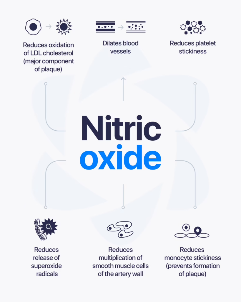
BPC-157 is a synthetic peptide that has been widely studied for its role in tissue repair and cellular signaling. One area that continues to draw attention is its potential interaction with nitric oxide, a molecule involved in blood flow and vascular function. Understanding this relationship helps researchers explore how peptides may influence key biological systems. What Is BPC-157? BPC-157 is a peptide derived from a protein found in gastric juice. In research settings, it is often studied for its connection to healing processes, including how tissues respond to stress or injury. Its stability and ability to interact with multiple pathways make it a frequent subject in peptide research. What Is Nitric Oxide? Nitric oxide is a signaling molecule produced naturally in the body. It plays a major role in regulating blood vessel function by helping vessels relax and expand. This process, known as vasodilation, allows for better blood flow and nutrient delivery throughout the body. How BPC-157 and Nitric Oxide May Be Connected Research suggests that BPC-157 may interact with pathways related to nitric oxide production and regulation. Some studies indicate that it may: Influence nitric oxide signaling pathways Support vascular response mechanisms Affect blood flow at the tissue level Interact with processes involved in repair and regeneration These interactions are of interest because nitric oxide is closely tied to how the body responds to injury and maintains circulation. Areas of Research Interest Researchers explore this connection across several areas: Vascular FunctionStudying how nitric oxide pathways affect blood flow and how peptides may influence those responses. Tissue Repair ProcessesUnderstanding how improved circulation and signaling contribute to healing at the cellular level. Cellular Signaling MechanismsAnalyzing how different pathways interact and regulate biological responses. What Current Research Suggests Most findings come from preclinical and laboratory studies. These suggest that BPC-157 may interact with nitric oxide systems in ways that influence vascular and cellular responses. However, results vary depending on the model and conditions used in research. Limitations and Ongoing Study Here’s the honest picture. While there is growing interest in the connection between BPC-157 and nitric oxide, large-scale human research is still limited. Most available data comes from: Animal studies Cell-based experiments Controlled laboratory research More research is needed to fully understand how these interactions behave across different contexts. Final Thoughts BPC-157 and nitric oxide are both important in understanding how the body regulates blood flow and tissue response. Their potential interaction makes them a valuable focus in ongoing peptide research. At this stage, this area remains under active investigation, with future studies expected to provide deeper insights into how these systems work together.

Cagrilintide is a synthetic peptide analog that has gained attention in research for its interaction with pathways related to appetite regulation and metabolic signaling. It is designed to mimic the activity of a naturally occurring hormone called amylin. Because of this, it’s often studied in connection with how the body manages energy balance and food intake. What Is Cagrilintide? Cagrilintide is an analog of amylin, a hormone that is normally released alongside insulin. Amylin plays a role in regulating appetite, slowing gastric emptying, and influencing how the body processes nutrients. By mimicking this hormone, Cagrilintide is studied to understand how these same pathways can be influenced in a controlled research setting. How It Works In research, Cagrilintide is examined for how it interacts with receptors involved in appetite and metabolic control. Some of its studied effects include: Influencing satiety signals Slowing the rate at which food leaves the stomach Affecting energy intake behavior Interacting with metabolic regulation pathways Rather than acting directly on energy production, it works through signaling mechanisms that help regulate intake and processing. Why It’s Being Studied Cagrilintide has become a focus in research because it targets specific pathways related to metabolic balance. Researchers are particularly interested in: Appetite RegulationUnderstanding how signals in the brain and body control hunger and fullness. Energy BalanceStudying how intake and expenditure are regulated through hormonal signaling. Peptide-Based PathwaysExploring how synthetic analogs can replicate and modify natural hormone activity. What Research Suggests So Far Most findings come from controlled studies and early-stage research. These suggest that Cagrilintide can influence appetite-related pathways through its interaction with amylin receptors. However, outcomes can vary depending on study design, dosage, and experimental conditions. Limitations and Current Understanding Here’s the honest take. While research is ongoing, there is still limited long-term data available across broader populations. Much of the current understanding is based on: Early-stage clinical research Laboratory studies Controlled experimental settings Further research is needed to fully understand its long-term behavior and broader implications. Final Thoughts Cagrilintide is a synthetic peptide designed to mimic amylin, making it a key subject in research focused on appetite and metabolic signaling. Its targeted mechanism has made it an area of growing interest. At this stage, it remains an active field of study, with ongoing research continuing to explore its full potential and applications.
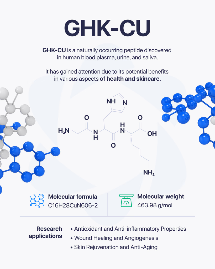
GHK-Cu is a naturally occurring peptide complex that has been widely studied for its role in cellular activity and tissue-related processes. It consists of a short peptide (GHK) bound to copper ions, which gives it unique properties in research settings. Because of this combination, it has become a point of interest in studies focused on cellular repair, signaling, and biological balance. What Is GHK-Cu Made Of? GHK-Cu is formed from a tripeptide made up of three amino acids: Glycine Histidine Lysine When this peptide binds with copper, it creates a complex that can interact with various biological systems. The copper component plays a key role in how the compound functions at the cellular level. How It Works in Research In research environments, GHK-Cu is studied for how it influences cellular communication and tissue-related processes. Some areas of focus include: Cellular repair mechanisms Regulation of gene expression Interaction with enzymes and proteins Support of structural components in tissues Its ability to bind copper allows it to participate in processes where metal ions are involved in biological activity. Why It’s Studied GHK-Cu has drawn attention because of its potential to influence multiple pathways at once. Researchers are interested in how it may support: Tissue-Related ProcessesStudying how cells repair and maintain structure. Cellular SignalingUnderstanding how signals are transmitted between cells. Structural Protein InteractionAnalyzing how it may affect components like collagen and other proteins. What Research Suggests Most available research comes from laboratory and preclinical studies. These suggest that GHK-Cu may play a role in regulating cellular processes and supporting tissue-related functions. However, outcomes can vary depending on conditions such as concentration, environment, and study design. Limitations and Current Understanding Here’s the honest picture. While GHK-Cu has been studied for many years, there is still ongoing research to fully understand all of its mechanisms and long-term effects. Most findings are based on: Cell-based studies Animal research Controlled laboratory environments More extensive research is needed to expand current knowledge. Final Thoughts GHK-Cu is a well-known peptide complex in research due to its connection to copper and its involvement in cellular processes. Its ability to interact with multiple biological pathways makes it an important subject of ongoing study. At this stage, it remains a key area of interest in peptide research, with continued investigation helping to uncover its full range of activity.

By Dr. Leonard Haberman, Chief Science Officer, OPTMZ Peptides | Published: April 2, 2026 | Last updated: April 17, 2026 In vitro research is performed outside a living organism — in controlled environments like cell cultures, isolated tissue preparations, or purified molecular systems. In vivo research is performed inside a living organism, such as an animal model. In vitro studies isolate a single variable under highly controlled conditions; in vivo studies observe how a compound behaves within a complete biological system. Both methods serve distinct scientific purposes, and rigorous preclinical peptide research typically progresses from one to the other rather than choosing between them. The terms themselves come from Latin: in vitro translates roughly to “in glass,” reflecting the historical use of test tubes and petri dishes, while in vivo translates to “in the living.” In modern research, the distinction is methodological rather than literal — in vitro systems now include sophisticated organ-on-chip platforms and 3D organoids, while in vivo encompasses everything from zebrafish models to non-human primate studies. What Does In Vitro Mean in a Research Context? An in vitro experiment takes place in a controlled artificial environment outside a living organism. Researchers use in vitro systems to study specific biological interactions in isolation — free from the confounding variables present in a whole organism. The goal is mechanistic precision. Common in vitro systems in peptide research include: Cell culture — immortalized cell lines (e.g., HEK293, HepG2, C2C12) or primary cell preparations maintained in growth media Receptor binding assays — isolated or membrane-bound receptor preparations used to characterize peptide-receptor affinity and selectivity Enzyme kinetics assays — purified enzyme systems used to measure substrate turnover or inhibition 3D organoid and spheroid cultures — multi-cellular constructs that more closely mimic tissue-level behavior Organ-on-chip platforms — microfluidic devices that recreate organ-level physiology in vitro In peptide research, in vitro assays are often the first experimental step after a compound has been synthesized and characterized. For example, studies investigating the tripeptide GHK-Cu have used in vitro fibroblast cultures to examine its interactions with collagen gene expression pathways (Pickart & Margolina, 2018 — PubMed). What Does In Vivo Mean in a Research Context? In vivo research is conducted inside a living organism. Rather than isolating one variable, in vivo studies observe how a compound interacts with the full biological context of a functioning system — circulation, metabolism, organ crosstalk, immune response, and tissue distribution. Common in vivo model systems include: Rodent models — mice and rats, used for the majority of preclinical pharmacology research Zebrafish — increasingly used for early-stage developmental and cardiovascular studies Non-human primates — reserved for late-stage preclinical work where physiological similarity to humans is essential Ex vivo preparations — tissues removed from a living organism and studied shortly after, occupying a middle category between in vitro and in vivo Peptide research frequently progresses from in vitro characterization into in vivo rodent work. BPC-157, a synthetic pentadecapeptide derived from a protective protein found in gastric juice, has been studied in rodent models for its effects on connective tissue healing kinetics (Chang et al., 2011 — PubMed; Seiwerth et al., 2018 — PubMed). These rodent studies followed earlier in vitro work characterizing the peptide’s interactions at the cellular level. How Do In Vitro and In Vivo Studies Fit in the Preclinical Research Progression? In modern peptide research, in vitro and in vivo approaches are rarely an either/or choice. They occupy different stages of a staged research progression: In silico — computational modeling of peptide structure, receptor docking, and predicted binding affinities. This stage requires no physical compound and is used to prioritize candidates for synthesis. In vitro — characterized peptide is tested in purified or cell-based systems to establish mechanism, selectivity, and dose-response behavior in isolation. Ex vivo — excised tissue or organ preparations bridge the gap between cell-based in vitro work and whole-organism in vivo work. In vivo — compound is evaluated in living animal models to assess pharmacokinetics, tissue distribution, and system-level effects. Clinical research — where applicable, and only after extensive preclinical work, compounds advance into human clinical trials under regulatory oversight. Each stage answers questions the previous stage could not. In vitro assays can demonstrate that a peptide binds its intended receptor with nanomolar affinity — but only in vivo work can show how that binding translates into distribution, metabolism, and physiological outcomes in a whole organism. In vivo work, in turn, cannot substitute for the mechanistic clarity that in vitro work provides. What Are the Strengths and Limitations of Each Approach? Each methodology offers distinct advantages and carries distinct constraints. The table below summarizes how in vitro and in vivo approaches compare across the dimensions researchers weigh when designing a preclinical study. Dimension In Vitro In Vivo What it measures Isolated mechanism under controlled conditions Compound behavior in a complete biological system Variable control High — single receptor, cell type, or pathway studied in isolation Low — biological complexity introduces many confounding factors Throughput High — large compound libraries screened in parallel Low — labor-intensive per subject Cost per data point Lower Higher Reproducibility High — controlled conditions yield consistent results Lower — biological variability between subjects Biological complexity Reduced — lacks tissue architecture, vascular supply, cross-system signaling Full — captures immune, endocrine, and metabolic integration Pharmacokinetics (ADME) Not captured Captured — absorption, distribution, metabolism, excretion Translational accuracy Indirect — in vitro potency often does not correspond to in vivo activity Closer to physiological reality; species differences remain Animal use None for primary screening Required, with ethical oversight Typical stage of use Early mechanistic characterization and screening Later-stage validation and system-level evaluation The methodological tradeoff: in vitro studies tell you what a compound can do; in vivo studies tell you what it does under real biological conditions. How Is In Vitro and In Vivo Research Applied to Peptide Compounds? Peptide compounds present particular methodological considerations across the in vitro / in vivo continuum. Stability and half-life. Many peptides degrade rapidly under physiological conditions. In vitro
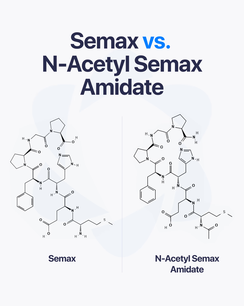
Semax and N-Acetyl Semax Amidate are closely related peptides often studied for their effects on the brain and cognitive processes. While they share a similar foundation, small structural differences between them can influence how they behave in research settings. Let’s break down what sets them apart. What Is Semax? Semax is a synthetic peptide derived from a fragment of adrenocorticotropic hormone (ACTH). It has been studied for how it interacts with brain signaling pathways, particularly those linked to cognitive function and neurotransmitter activity. In research, Semax is often explored for its potential influence on: Cognitive processes such as memory and focus Neurotransmitter regulation Brain-derived signaling pathways What Is N-Acetyl Semax Amidate? N-Acetyl Semax Amidate is a modified version of Semax. It includes two structural changes: An added acetyl group An amidated end These modifications are designed to improve stability and resistance to breakdown, allowing the peptide to remain active longer in research conditions. Key Differences Between the Two Here’s the simple comparison: Semax is the original peptide structure N-Acetyl Semax Amidate is a more stable, modified version Because of its modifications, N-Acetyl Semax Amidate is often studied for: Increased stability Longer duration in experimental conditions Potentially more consistent activity over time Why Stability Matters Peptides can degrade quickly depending on environmental conditions. By modifying the structure, researchers can observe how increased stability affects performance and interaction with biological systems. This is one of the main reasons analog versions like N-Acetyl Semax Amidate are developed and studied. Areas of Research Interest Both peptides are explored in similar research areas: Cognitive FunctionStudying how they may influence memory, learning, and focus-related pathways. Neurotransmitter ActivityAnalyzing interactions with systems such as dopamine and serotonin. Brain Signaling MechanismsUnderstanding how peptide-based compounds affect neural communication. What Research Suggests Most findings come from laboratory and early-stage studies. These indicate that both compounds interact with neurological pathways, but differences in stability may affect how long and how consistently they act. However, results can vary based on experimental conditions. Limitations and Ongoing Research Here’s the honest take. While both peptides are actively studied, there is limited large-scale clinical data comparing them directly. Most research is based on: Preclinical studies Controlled lab experiments Smaller-scale investigations More research is needed to fully understand how these differences translate across broader contexts. Final Thoughts Semax and N-Acetyl Semax Amidate are closely related peptides with similar research focus areas. The main difference lies in structural modifications that may influence stability and duration. For researchers, choosing between them often comes down to whether stability and extended activity are important for the specific study.
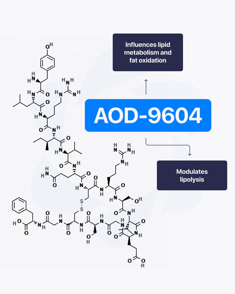
By Dr. Leonard Haberman, Chief Science Officer, OPTMZ Peptides Published April 2, 2026 | Last updated April 16, 2026 Research Use Only. All products and information referenced in this article are intended for laboratory research conducted by qualified professionals. AOD-9604 is not approved by the FDA for any clinical use and is not intended for human or animal consumption. Statements in this article have not been evaluated by the FDA and are not intended to diagnose, treat, cure, or prevent any disease. AOD-9604 is a synthetic 16-amino-acid peptide corresponding to the C-terminal fragment (residues 176–191) of human growth hormone (hGH). It was developed in the late 1990s by Metabolic Pharmaceuticals (Australia) and has been investigated in pre-clinical metabolic research and, more recently, in tissue and cartilage research models. AOD-9604 is sold for laboratory research use only; it is not an FDA-approved therapeutic agent. What Is AOD-9604? AOD-9604 is a hexadecapeptide engineered to replicate a specific functional segment of hGH — the lipolytic domain — without reproducing the full hormone’s growth-promoting or insulin-modulating activity. Researchers refer to the same molecule by several names: AOD-9604, AOD9604, hGH fragment 176-191, and (less commonly in current literature) hGH 177-191. The compound carries CAS Number 221231-10-3, a molecular formula of C₇₈H₁₂₃N₂₃O₂₃S₂, and a molecular weight of approximately 1,815.1 g/mol. Its PubChem CID is 71300630. The molecule was synthesized to investigate whether the lipid-pathway-related activity of growth hormone could be isolated from its other endocrine effects. In animal model research conducted at Monash University and by Metabolic Pharmaceuticals, AOD-9604 was reported to induce changes in lipid metabolism without producing the IGF-1 elevation, glucose effects, or growth signaling associated with full-length hGH (Ng et al., 2000) PubMed: 11146367. AOD-9604 Molecular Structure and Origin: The HGH Fragment 176-191 The full hGH molecule contains 191 amino acids. Early structure-activity research conducted in the 1980s and 1990s identified the C-terminal region — specifically residues 177–191, with a tyrosine added at position 176 to produce a stable hexadecapeptide — as the segment most associated with measurable lipid-pathway effects in vitro and in animal models. Metabolic Pharmaceuticals patented the resulting analogue, designated AOD-9604, in the late 1990s and advanced it through a series of pre-clinical and Phase I/II investigations between 2001 and 2007 (Stier et al., 2013) jofem.org. A 2010 review of obesity pharmacology characterized AOD-9604 as an orally and parenterally bioavailable analogue of the hGH 177-191 region that demonstrated lipolytic activity in animal trials but produced limited efficacy signals in later-stage human studies (Isidro & Cordido, 2010). The compound’s clinical development as a therapeutic was discontinued in 2007 following Phase 2B trial outcomes that did not support commercial advancement (Valentino et al., 2010) PMC3136748. For research-grade verification, the molecular structure is commonly confirmed by mass spectrometry (target m/z corresponding to the 1,815.1 g/mol molecular weight) and purity is quantified by reversed-phase HPLC against a reference standard. These methods, along with endotoxin and microbial testing, comprise the standard analytical panel applied to AOD-9604 batches sold for research use. How Does AOD-9604 Work in Research Models? The most-studied biochemical activity of AOD-9604 in pre-clinical models involves interaction with lipid-metabolism pathways in adipose tissue. Heffernan and colleagues, working at Monash University, examined chronic AOD-9604 administration in obese mice and in β3-adrenergic receptor knock-out mice. Their 2001 paper in Endocrinology reported that the compound produced effects on lipolytic sensitivity that appeared to depend at least partially on β3-adrenergic receptor signaling — a pathway distinct from the GH receptor — and did not measurably stimulate IGF-1 production (Heffernan et al., 2001) PubMed: 11713213. A separate study by Ng and colleagues in Hormone Research examined AOD-9604 in obese Zucker rats, a standard rodent model in metabolic research, and reported observations consistent with isolated lipid-pathway activity in the absence of measurable somatogenic effects (Ng et al., 2000) PubMed: 11146367. Together, these foundational papers established the research framework in which AOD-9604 has subsequently been investigated. It is important to note that the mechanism described in animal models has not translated into clinically meaningful human efficacy. The Phase 2B human trial conducted by Metabolic Pharmaceuticals between 2004 and 2007 did not produce results sufficient to support continued development, and AOD-9604 has not been approved by the FDA, EMA, or TGA for any therapeutic indication. What Have Published Studies of AOD-9604 Examined? The published literature on AOD-9604 spans roughly 25 years and includes pre-clinical animal studies, human safety trials, and more recent investigations in tissue research contexts. Pre-clinical metabolic research The earliest characterizations of AOD-9604 focused on lipid-pathway interactions in rodent models. Ng et al. (2000) and Heffernan et al. (2001) remain the most-cited references for the foundational pharmacology, and both papers are openly accessible through PubMed. Human safety and tolerability research Stier and colleagues published a 2013 review in the Journal of Endocrinology, Metabolism and Diabetes of South Africa summarizing six human clinical trials conducted between 2001 and 2006 involving 893 participants. The review reported that AOD-9604 was generally well-tolerated in short-term administration and did not produce clinically significant changes in IGF-1, glucose, or insulin parameters compared to placebo (Stier et al., 2013) jofem.org. A subsequent 2014 publication by Moré and colleagues reviewed long-term safety and genotoxicology data and reported no evidence of mutagenic or toxicological concerns within the parameters studied (Moré et al., 2014). Cartilage and tissue research More recent literature has examined AOD-9604 in the context of cartilage and connective tissue research. A 2026 review of therapeutic peptides in orthopaedic research models discussed AOD-9604 alongside other peptide fragments studied for their potential interactions with chondrocyte signaling and tissue maintenance pathways (Rahman et al., 2026) PMC12753158. This research direction remains preliminary, with most published work confined to in vitro and small-animal models. Across all three research contexts, the published data is best characterized as exploratory. Sample sizes are limited, the body of human evidence is small, and no large-scale efficacy trials have been completed for any indication. What Is AOD-9604’s Regulatory Status? AOD-9604 is not approved by the U.S. Food and Drug Administration for any clinical use. It
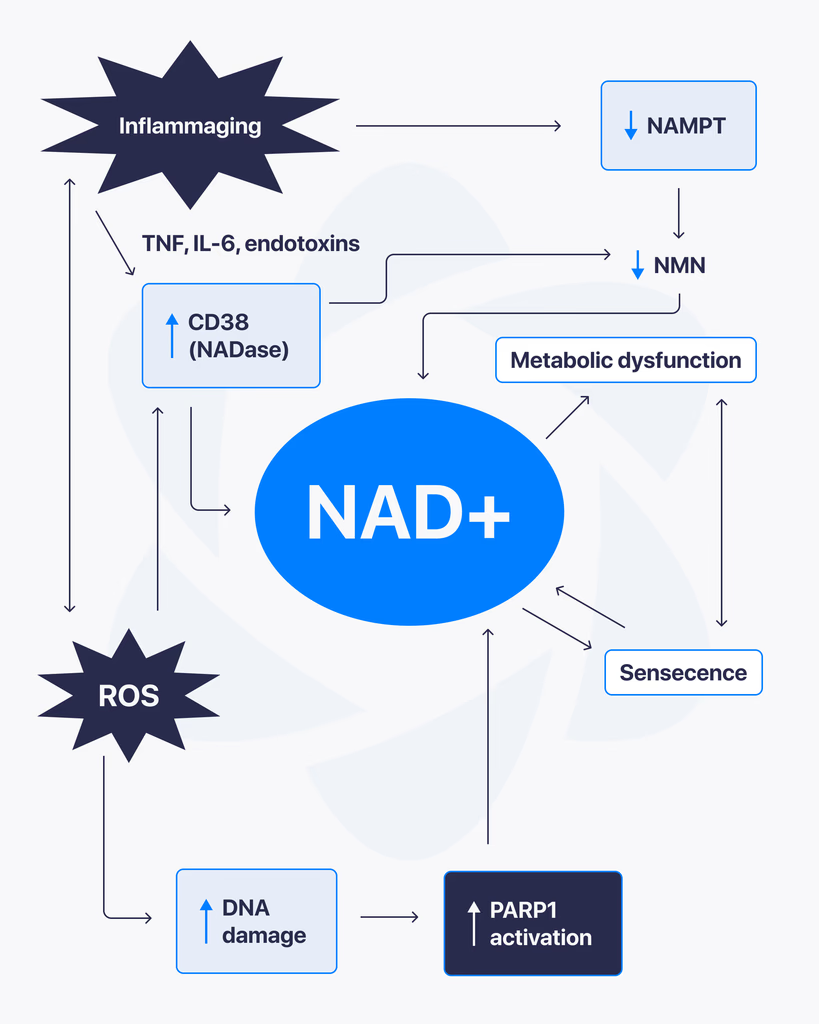
NAD+, short for nicotinamide adenine dinucleotide, is a coenzyme found in every living cell. It plays a central role in energy production and cellular function, which is why it has become a major focus in ongoing research. Scientists are studying how NAD+ levels influence different biological processes, especially those related to metabolism and cellular health. What Is NAD+? NAD+ is a molecule that helps transfer energy within cells. It’s involved in reactions that convert nutrients into usable cellular energy. As cells perform their functions, NAD+ cycles between two forms, helping drive essential processes that keep cells active and functioning. Why NAD+ Matters NAD+ is critical for several core cellular functions, including: Energy production within mitochondria DNA repair mechanisms Regulation of cellular stress responses Support of metabolic processes Because of its wide-ranging role, changes in NAD+ levels can affect how efficiently cells operate. NAD+ and Cellular Energy One of the main reasons NAD+ is studied is its connection to energy metabolism. It helps facilitate reactions that produce ATP, the primary energy source used by cells. Without sufficient NAD+, these processes become less efficient, which can impact overall cellular performance. What Research Is Exploring Researchers are examining how NAD+ interacts with different pathways inside the body. Some key areas of focus include: Mitochondrial FunctionUnderstanding how NAD+ supports energy production and efficiency within cells. Cellular Repair SystemsStudying its role in activating enzymes involved in DNA repair and maintenance. Metabolic RegulationExploring how NAD+ levels influence how the body processes and uses energy. What Studies Suggest So Far Most findings come from laboratory and preclinical research. These studies indicate that NAD+ plays a vital role in maintaining cellular function and responding to stress. There is also growing interest in how NAD+ levels change over time and how that may affect different biological systems. Limitations and Current Understanding Here’s the reality. While NAD+ is well understood at a biochemical level, many of its broader effects are still being researched. Most available data comes from: Cell-based studies Animal research Early-stage human investigations More comprehensive research is needed to fully understand long-term impacts and practical applications. Final Thoughts NAD+ is a key molecule in cellular energy and maintenance, making it an important subject in modern research. Its involvement in multiple biological pathways continues to drive interest across different scientific fields. At this stage, it remains an active area of study, with ongoing research working to better understand how it influences cellular function over time.

Acetic acid is a widely used compound in chemical and laboratory settings, playing a role in everything from synthesis processes to analytical applications. As research continues to evolve, new methods are improving how acetic acid is produced, making the process more efficient, scalable, and environmentally conscious. What Is Acetic Acid? Acetic acid is a simple organic compound known for its role in various chemical reactions. In research and industrial settings, it’s commonly used as a solvent, reagent, and intermediate in synthesis. Its versatility makes it an important component in both small-scale laboratory work and large-scale production systems. Traditional Production Methods Historically, acetic acid has been produced through processes such as: Methanol carbonylation Oxidation of acetaldehyde Fermentation-based methods Among these, methanol carbonylation has become the dominant industrial approach due to its efficiency and scalability. What’s Changing in 2025 Recent developments are focused on improving sustainability, reducing waste, and increasing precision in production. Advanced CatalystsNew catalyst systems are being developed to improve reaction efficiency and reduce energy consumption. These catalysts help speed up production while minimizing unwanted byproducts. Greener Production TechniquesThere is a growing shift toward environmentally friendly methods, including bio-based production and reduced reliance on harsh chemicals. Process OptimizationModern systems are using improved monitoring and control technologies to ensure more consistent output and better resource management. Integration with Renewable InputsSome research is exploring the use of renewable feedstocks, aiming to reduce dependence on traditional petrochemical sources. Why These Innovations Matter Improving how acetic acid is produced has a direct impact on research and manufacturing. Better processes mean: Higher purity and consistency Reduced environmental impact Lower production costs over time More reliable supply chains For laboratories and industries that rely on precise chemical inputs, these improvements make a noticeable difference. Applications in Research Acetic acid continues to be used across various research areas, including: Chemical synthesis Analytical testing Peptide and organic compound preparation Buffer and solvent systems Its role as a foundational compound ensures it remains relevant as research techniques advance. Final Thoughts Acetic acid synthesis is evolving with a clear focus on efficiency and sustainability. Innovations in catalysts, production methods, and process control are shaping how this essential compound is produced and used. As research continues, these advancements are expected to further improve both quality and environmental impact, making acetic acid production more refined and future-ready.
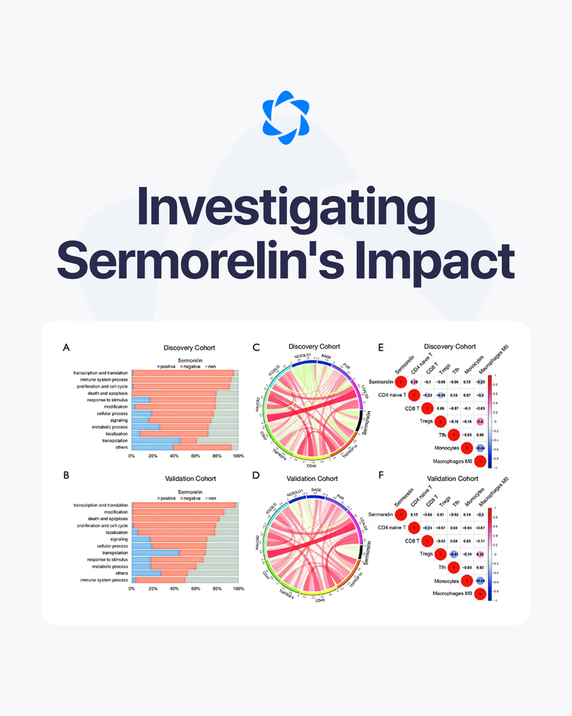
By Dr. Leonard Haberman, Chief Science Officer, OPTMZ Peptides Published: April 2, 2026 · Last updated: April 16, 2026 Sermorelin is a 29-amino-acid synthetic peptide corresponding to residues 1–29 of growth hormone-releasing hormone (GHRH 1–44), the biologically active N-terminal fragment. In research models, sermorelin binds the GHRH receptor on anterior pituitary somatotrophs, activates adenylate cyclase, and triggers a cyclic AMP–protein kinase A (cAMP–PKA) signaling cascade that has been studied for its role in growth hormone gene transcription and release (Walker, 2006). Investigators have used sermorelin to examine GHRH-receptor pharmacology, downstream second-messenger dynamics, and — more recently — its interactions with cell proliferation pathways in oncology research models (Chang et al., 2021). This research overview summarizes what controlled studies have documented about sermorelin’s molecular mechanism, its half-life behavior in research contexts, what cell proliferation data has emerged, and how research-grade material is verified for laboratory use. What Is Sermorelin? Sermorelin is the truncated 1–29 amino acid sequence of native human GHRH, retaining the receptor-binding domain required for GHRH-R activation. The full-length endogenous peptide is 44 amino acids, but research has established that the first 29 residues are sufficient to reproduce the agonist activity of the parent molecule (Walker, 2006). The compound is supplied for research as a lyophilized powder requiring reconstitution in bacteriostatic water before use in cell-based or in-vivo research protocols. Structurally, sermorelin is classified as a GHRH analog rather than a growth hormone analog — a distinction that matters for experimental design. It does not interact directly with growth hormone receptors. Instead, it acts upstream at the pituitary level, making it a tool for studying GHRH-R signaling rather than peripheral GH-receptor activity. How Does Sermorelin Work? The GHRH Receptor Mechanism The mechanism of action observed in pre-clinical research follows a defined sequence at the molecular level: Receptor binding. Sermorelin binds to the GHRH receptor (GHRH-R), a class B G-protein-coupled receptor (GPCR) expressed on somatotroph cells of the anterior pituitary. G-protein coupling. Receptor activation triggers conformational changes that activate the Gαs subunit of the associated heterotrimeric G-protein. Adenylate cyclase activation. Gαs stimulates membrane-bound adenylate cyclase, which catalyzes the conversion of ATP to cyclic AMP (cAMP). PKA cascade. Elevated intracellular cAMP activates protein kinase A (PKA), which phosphorylates target proteins including the transcription factor CREB. Transcriptional regulation. Phosphorylated CREB binds the GH gene promoter, increasing transcription of growth hormone messenger RNA. Walker (2006) noted that this pathway “stimulates pituitary gene transcription of hGH messenger RNA, increasing pituitary reserve.” This signaling architecture is what makes sermorelin useful in research: it engages a well-characterized GPCR pathway with measurable second-messenger readouts (cAMP accumulation assays, CREB phosphorylation Western blots, GH ELISA from culture supernatant). A practical consequence observed in research contexts is that GH release driven by sermorelin remains under physiological feedback control by somatostatin and IGF-1, producing pulsatile rather than tonic patterns in animal models — a behavior distinct from exogenous recombinant GH administration (Walker, 2006). What Is the Half-Life of Sermorelin in Research Models? Sermorelin has a notably short plasma half-life — published research data places it at approximately 11–12 minutes in circulation, which is characteristic of GHRH-family peptides and reflects rapid enzymatic degradation by dipeptidyl peptidase-IV (DPP-IV) and other peptidases (Walker, 2006). For research workflows, this short half-life has several implications: Sampling windows are narrow. Time-course experiments measuring GH release require closely spaced sampling intervals (typically every 5–15 minutes for the first hour). Storage and reconstitution discipline matters. Once reconstituted, sermorelin should be maintained at 2–8°C and used within the verified stability window indicated on the batch certificate of analysis. Comparison studies use longer-acting analogs. Researchers comparing pulsatile versus sustained GHRH-R activation often pair sermorelin against modified GHRH analogs with extended half-lives (CJC-1295 and modified GRF 1–29 derivatives). What Has Research Shown About Sermorelin and Cell Proliferation? The most cited contemporary research on sermorelin and cellular proliferation is Chang et al. (2021), which used bioinformatic analysis (gene ontology and KEGG pathway enrichment) to examine sermorelin’s potential as a candidate for recurrent glioma research. The investigators reported that gene ontology analysis identified sermorelin as “closely related to cell proliferation functions” and proposed that the compound may inhibit tumor cell proliferation through cell cycle blocking mechanisms. Independent supporting work by Muñoz-Moreno et al. (2018) investigated GHRH-receptor antagonists in LNCaP and PC3 prostate cancer cell lines. While that work studied antagonists rather than agonists, it established that the GHRH-R signaling axis is functionally engaged in proliferative cell models — providing the receptor-pathway context that makes sermorelin’s effects in Chang’s bioinformatic analysis biologically plausible. It is important to characterize this body of work accurately: the cell proliferation data is bioinformatic and pre-clinical. There are no large-scale human studies investigating sermorelin’s effects on cellular proliferation. Researchers working in this area are typically running cell-line experiments, animal models, or computational analyses — not clinical trials. For laboratory teams designing experiments in this space, the key methodological considerations are: receptor expression validation in the chosen cell line (GHRH-R is not expressed at meaningful levels in all immortalized lines), batch-to-batch purity verification of the sermorelin used (impurities or degradation products can confound proliferation readouts), and inclusion of appropriate controls for cAMP-PKA pathway activation. How Does Sermorelin Compare to Other GHRH-Related Research Peptides? Within the GHRH-analog family, sermorelin is distinguished by its short half-life and its identity as the unmodified 1–29 sequence. Researchers comparing GHRH-pathway tools typically evaluate: CJC-1295 — a modified GRF 1–29 with a drug affinity complex (DAC) extension that significantly extends plasma half-life, used in research where sustained receptor engagement is the experimental variable. Tesamorelin — a stabilized GHRH 1–44 analog with an N-terminal trans-3-hexenoyl modification that resists DPP-IV cleavage. Ipamorelin — not a GHRH analog at all, but a ghrelin-receptor (GHS-R) agonist that converges on the same downstream output (GH release) through a parallel receptor system. This makes ipamorelin a useful comparator in studies dissecting the contributions of the two upstream pathways. The choice between these tools depends on the experimental question. Sermorelin’s short half-life and unmodified sequence make it the cleanest tool for studying acute,