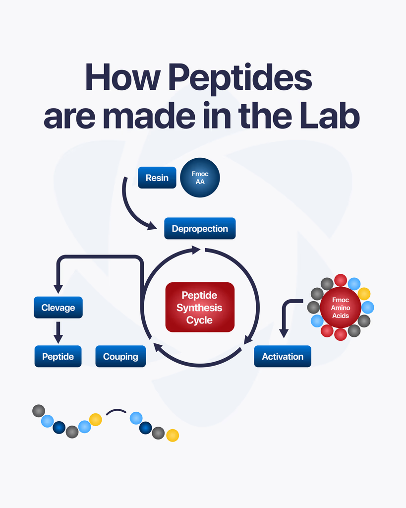
By Dr. Leonard Haberman, Chief Science Officer, OPTMZ Peptides Published: March 2, 2026 | Last Updated: April 16, 2026 Peptide synthesis is the controlled laboratory process by which researchers build peptide chains from individual amino acid subunits. Rather than isolating peptides from biological tissues, synthetic production allows precise sequence specification — making it the standard method for producing research-grade compounds used in pre-clinical and in vitro studies. Understanding how research peptides are synthesized helps researchers make better decisions about supplier selection, purity interpretation, and storage protocols. This article covers the core synthesis method, the purification steps that follow, and the analytical testing that determines whether a finished batch meets research-grade standards. What Is Peptide Synthesis? Peptide synthesis is the process of forming peptide bonds between amino acids in a defined sequence. In biological systems, this occurs through ribosomal translation of mRNA sequences. In laboratory settings, synthesis is performed chemically — allowing researchers to specify any sequence, introduce non-natural amino acids, or produce compounds that do not exist in sufficient quantities in natural sources. The peptide bond itself is a covalent amide linkage between the carboxyl group of one amino acid and the amino group of the next. Each addition extends the chain by one residue. A ten-residue peptide requires nine successful bond formations, each carried out under controlled conditions (Merrifield, 1963 — PubMed). The precision of the final sequence determines the compound’s research relevance. A single incorrect residue, deleted residue, or epimeric impurity can alter receptor binding, biological stability, or assay behavior. This is why synthesis methodology and post-synthesis verification are inseparable from research utility. What Is Solid-Phase Peptide Synthesis (SPPS)? Solid-phase peptide synthesis (SPPS) is the dominant method for producing research-grade peptides. Introduced by Robert Bruce Merrifield in 1963, the technique anchors the growing peptide chain to an insoluble polymer resin, which allows excess reagents and byproducts to be washed away between each coupling step (Merrifield, 1963 — PubMed). How Does SPPS Work? The synthesis proceeds through a repeating cycle: 1. Resin loading. The C-terminal amino acid of the target sequence is attached to the solid resin support via a linker. The resin physically anchors the chain throughout synthesis, enabling filtration-based purification between steps. 2. Deprotection. Amino acids carry protecting groups on their reactive side chains and N-terminus. Before each coupling step, the N-terminal protecting group is removed — a process called deprotection — exposing the free amino group for the next reaction. 3. Coupling. The next amino acid in the sequence is added in excess with a coupling reagent. The coupling reagent activates the incoming amino acid’s carboxyl group, enabling it to react with the deprotected N-terminus on the resin-bound chain. This forms a new peptide bond. 4. Washing. Excess reagents, activating agents, and byproducts are washed away with solvent. The resin retains the growing peptide chain. 5. Iteration. The deprotection → coupling → washing cycle repeats for each amino acid in the sequence, proceeding from C-terminus to N-terminus. 6. Cleavage and global deprotection. When the full sequence has been assembled, the completed peptide is cleaved from the resin. Simultaneously, remaining side-chain protecting groups are removed, yielding the free peptide in solution. The primary advantage of SPPS over solution-phase synthesis is the ability to use large excesses of coupling reagents to drive reactions to completion — a requirement when even a small percentage of incomplete couplings results in deletion sequences or truncated impurities that reduce the final purity. Two main SPPS strategies exist based on the N-terminal protecting group chemistry used: Boc (t-Boc) SPPS and Fmoc SPPS. Fmoc chemistry has become the standard for most research peptide synthesis due to its milder deprotection conditions and compatibility with a wider range of side-chain protecting groups (Carpino and Han, 1972 — PubMed). Why Do Incomplete Coupling Reactions Matter for Research? Each coupling step in SPPS does not achieve 100% efficiency. A typical coupling reaction yields 98–99.9% efficiency depending on the amino acid, the sequence context, and the coupling reagent used. Across a long sequence, these small inefficiencies accumulate. For a 20-residue peptide synthesized at 99% per-step coupling efficiency, the theoretical yield of the fully correct sequence is approximately 82% (0.99²⁰). The remaining fraction consists of deletion peptides — sequences where one or more amino acids have been skipped — and truncated sequences that terminated early. These impurities do not always share the same biological behavior as the target peptide. In research applications where receptor selectivity, binding affinity, or downstream signaling effects are being studied, impurity profiles that deviate significantly from the intended sequence can introduce confounding variables into experimental results (Chan and White, 2000 — PubMed). This is why purification is not optional for research-grade compounds. How Is a Research Peptide Purified After Synthesis? The crude peptide cleaved from the resin contains the target compound alongside synthesis byproducts, incomplete sequences, and protecting group fragments. Purification removes these impurities to yield a product that meets defined purity thresholds for research use. Reversed-Phase High-Performance Liquid Chromatography (RP-HPLC) Reversed-phase HPLC (RP-HPLC) is the standard purification method for research peptides. The crude peptide mixture is dissolved and injected onto an HPLC column packed with a hydrophobic stationary phase (typically C18). As a mobile phase gradient of water and organic solvent (usually acetonitrile) moves through the column, peptide components separate based on their hydrophobicity. The target peptide elutes at a characteristic retention time, and the corresponding fraction is collected. Other fractions — containing deletion sequences, oxidation products, and protecting group adducts — are discarded. Preparative RP-HPLC can achieve purity levels exceeding 98% for most research peptide sequences. The resulting material is lyophilized (freeze-dried) to produce a stable white to off-white powder suitable for storage and research use. How Is Peptide Purity Analytically Verified? Purification produces a compound of defined purity. Analytical testing confirms that purity and verifies the compound’s identity. For research-grade peptides, this is not a quality assurance formality — it is the primary evidence that the compound in the vial is what the label states. What Does HPLC Purity Testing Measure? Analytical
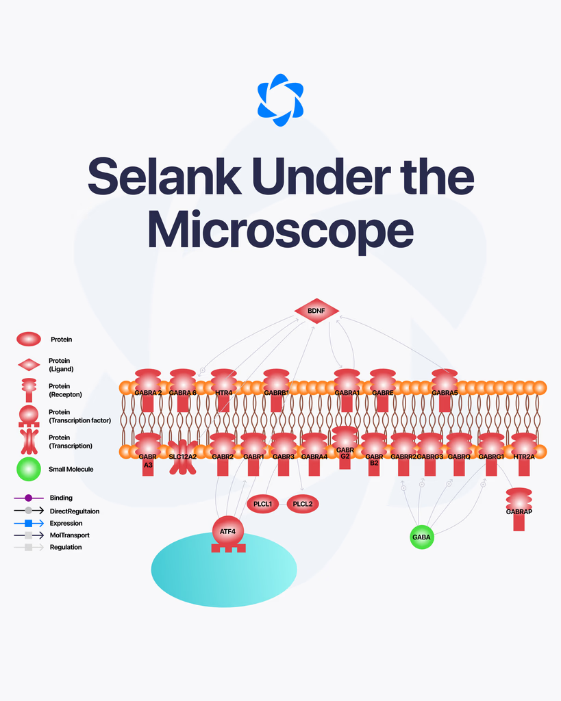
Selank is a synthetic peptide that has been studied for its potential effects on the nervous system. It’s derived from a naturally occurring peptide involved in immune signaling, but modified to improve stability and activity in research settings. Over time, it has gained attention for how it may interact with brain function, particularly in areas related to mood and cognitive processes. What Is Selank? Selank is a short peptide analog based on tuftsin, a naturally occurring compound linked to immune system activity. Researchers have explored how this modified version behaves in the body, especially in relation to neurological pathways. Because of its structure, Selank is often studied for its potential to influence signaling in the brain without acting like traditional compounds. How It’s Studied in Research In laboratory settings, Selank is examined for how it interacts with neurotransmitters and brain receptors. Some studies suggest it may affect systems involving: GABA (gamma-aminobutyric acid) Serotonin pathways Dopamine balance These systems are all closely tied to mood regulation, stress response, and cognitive function. Potential Areas of Interest Researchers have explored several areas where Selank may play a role: Cognitive FunctionSome studies suggest it may support memory and learning processes by influencing how signals are transmitted in the brain. Stress and Anxiety ResponseThere is interest in how Selank interacts with stress-related pathways, particularly through its effect on neurotransmitter balance. Immune System InteractionSince it is derived from tuftsin, Selank is also studied for how it may influence immune signaling and inflammatory responses. What Current Research Suggests Most findings around Selank come from preclinical and early-stage studies. These suggest that the peptide may have regulatory effects on certain brain functions without producing strong sedative or stimulant effects. However, it’s important to understand that research is still developing. There is limited large-scale clinical data, and many findings are based on controlled laboratory environments. Limitations and Ongoing Study Here’s the honest picture. While early research is promising, there isn’t enough conclusive evidence to fully define Selank’s role across all potential areas. Most studies focus on: Animal models Cellular-level analysis Small-scale research trials Further investigation is needed to better understand long-term effects and broader applications. Final Thoughts Selank is an interesting subject within peptide research, especially when it comes to studying brain function and signaling pathways. Its interaction with neurotransmitters and immune-related processes makes it a focus for ongoing research. For now, it remains a compound being actively studied rather than a fully established solution in any specific area.
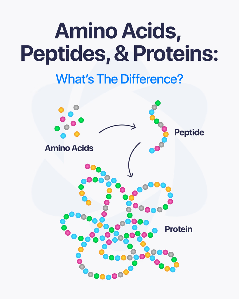
If you’re working around peptide research, you’ll keep seeing these three terms: amino acids, peptides, and proteins. They’re closely connected, but not the same thing. Understanding how they relate makes everything else easier to follow. Starting Point: Amino Acids Amino acids are the smallest units in this chain. They’re the basic building blocks that make up both peptides and proteins. Each amino acid has a simple structure, but when they link together, they form more complex compounds. There are many types, and the specific sequence in which they connect determines the final structure and function. In short, amino acids are the foundation. What Are Peptides? Peptides are short chains of amino acids linked together through peptide bonds. You can think of them as the middle layer between single amino acids and full proteins. They usually contain a limited number of amino acids, often ranging from just a few to a few dozen. Because of their smaller size, peptides are often easier to study and analyze in research settings. Peptides are involved in many biological processes, including signaling and regulation. What Are Proteins? Proteins are much larger and more complex structures made from long chains of amino acids. These chains fold into specific shapes, which determine how the protein functions. Unlike peptides, proteins can contain hundreds or even thousands of amino acids. This complexity allows them to perform a wide range of functions, from structural support to enzymatic activity. The Key Differences Here’s the simplest way to break it down: Amino acids are individual building blocks Peptides are short chains of amino acids Proteins are long, fully structured chains with complex functions The difference mainly comes down to size, structure, and biological role. Why This Matters in Research In peptide research, knowing where a compound fits in this chain helps researchers understand how it behaves. For example: Amino acids are studied for their chemical properties Peptides are often used to explore signaling and targeted interactions Proteins are analyzed for their broader biological functions Each level offers different insights depending on the goal of the research. Final Thoughts Amino acids, peptides, and proteins are all part of the same biological system, just at different levels of complexity. Once you understand how they connect, it becomes much easier to navigate research and terminology in this field.
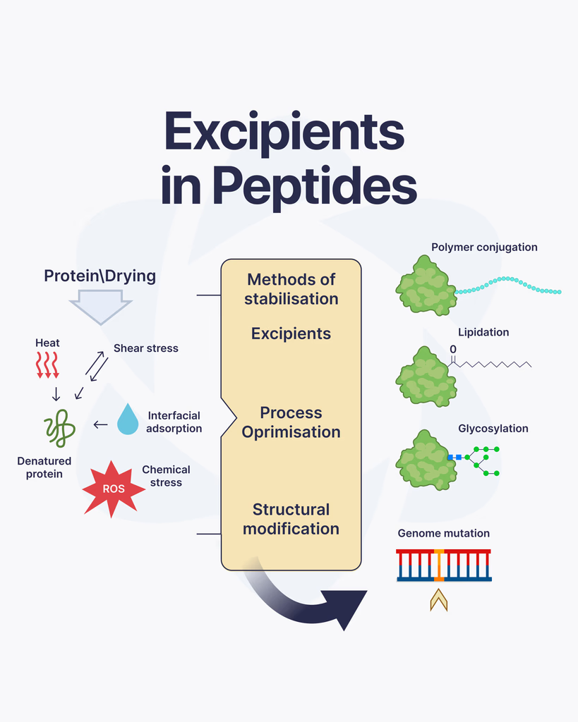
When working with peptides, most of the attention goes to the active compound itself. But there’s another important component that often gets overlooked, excipients. These supporting substances play a key role in how peptides are stored, handled, and studied. What Are Excipients? Excipients are inactive substances added alongside a peptide. They don’t produce biological effects on their own, but they help maintain the stability and usability of the peptide during storage and research. Think of them as support agents. Their job is to protect the peptide, improve consistency, and make handling easier in laboratory settings. Why Excipients Are Used in Peptide Research Peptides can be sensitive to environmental conditions like temperature, moisture, and light. Without proper support, they may degrade or lose effectiveness. Excipients are used to: Stabilize peptide structure Extend shelf life Prevent degradation during storage Improve solubility for reconstitution Ensure consistent handling across batches This makes them essential for maintaining reliability in research work. Common Types of Excipients Different excipients are selected depending on the peptide and how it will be used in research. Some of the most common include: Bulking AgentsThese help give the peptide substance physical volume, especially in lyophilized (freeze-dried) form. Mannitol is a commonly used example. StabilizersStabilizers protect peptides from breaking down over time. They help maintain the molecular structure during storage and transport. BuffersBuffers control pH levels, which is important because peptides can be sensitive to even small changes in acidity or alkalinity. SolubilizersThese help peptides dissolve properly when being prepared for research use, ensuring accurate concentration and handling. How Excipients Affect Research Quality Even though excipients are inactive, they still influence how a peptide performs in a research environment. Poor formulation can lead to instability, inaccurate dosing, or inconsistent results. That’s why high-quality peptide preparation always includes carefully selected excipients that match the compound’s characteristics. Things to Keep in Mind Not all excipients are the same, and their compatibility with specific peptides matters. Researchers often consider: The chemical stability of the peptide Storage conditions and shelf life Reconstitution requirements Interaction between excipients and the peptide Choosing the right combination is part of ensuring reliable and repeatable outcomes. Final Thoughts Excipients may not be the main focus in peptide research, but they play a critical supporting role. From stabilizing the compound to improving handling and storage, they help ensure that peptides remain consistent and usable throughout the research process. Ignoring excipients can lead to compromised results, while the right formulation helps maintain accuracy and confidence in every study.

By Dr. Leonard Haberman, Chief Science Officer, OPTMZ Peptides Published: March 2, 2026 | Last Updated: April 15, 2026 What Are Sermorelin and Ipamorelin? Sermorelin and ipamorelin are growth hormone (GH) secretagogues — compounds that stimulate GH release from anterior pituitary somatotroph cells through distinct receptor-mediated mechanisms. In peptide research, they serve as representative model compounds for two separate GH regulatory pathways: the GHRH receptor pathway (sermorelin) and the ghrelin receptor pathway (ipamorelin). Researchers studying GH axis regulation select between these compounds based on which receptor mechanism is under investigation — they are not functionally interchangeable. Both compounds are available from OPTMZ Peptides as batch-verified research reagents, independently tested by Krause Analytical — DEA-registered, ISO/IEC 17025-certified. Batch-level Certificates of Analysis are published at the COA Vault → Understanding the GH Axis: GH, GHRH, and GHRP Growth hormone regulation involves three primary elements that frequently appear together in secretagogue research. Growth Hormone (GH / Somatotropin) is a 191-amino acid peptide hormone secreted by anterior pituitary somatotrophs. In pre-clinical research, GH is studied for its downstream effects on IGF-1 axis activation, lipid metabolism signaling, and cellular anabolic pathways. It is the effector molecule — the end product of the regulatory cascade that GHRH and GHRPs each modulate. Growth Hormone Releasing Hormone (GHRH) is a 44-amino acid neuropeptide produced in the arcuate nucleus of the hypothalamus. GHRH acts on the GHRH receptor (GHRHR) expressed on somatotroph cells, activating Gαs-coupled adenylyl cyclase, increasing intracellular cAMP, and driving GH synthesis and pulsatile secretion. Sermorelin is a synthetic GHRH analog that targets this pathway. Growth Hormone Releasing Peptides (GHRPs) are a structurally distinct class of synthetic secretagogues that stimulate GH release through a separate receptor — the GH secretagogue receptor type 1a (GHS-R1a), also known as the ghrelin receptor. GHRPs were first characterized in binding studies by Bowers CY in the 1980s–1990s and represent an independent intervention point for GH axis research (Bowers CY, Cell Mol Life Sci, 1998). Ipamorelin is the most receptor-selective GHRP studied to date. Research has demonstrated that simultaneous stimulation of both the GHRHR and GHS-R1a pathways produces synergistic GH release exceeding the additive effect of either compound alone — a finding that has made dual-pathway co-administration a standard experimental model in neuroendocrinology research (Sigalos JT, Pastuszak AW, Sex Med Rev, 2018). Sermorelin: GHRH Receptor Agonist in Research Molecular Structure and Receptor Mechanism Sermorelin is the N-terminal 29-amino acid fragment of native human GHRH — designated hGHRH[1-29]NH₂. This truncated fragment retains full biological activity at the GHRHR. The C-terminal residues beyond position 29 contribute to plasma stability but are not required for receptor binding or activation. The receptor mechanism proceeds as follows: sermorelin binds the extracellular domain of GHRHR on anterior pituitary somatotrophs, activating the Gαs protein-coupled adenylyl cyclase system. This increases intracellular cAMP, activating protein kinase A (PKA), which drives both acute GH exocytosis and GH gene transcription. The result is GH secretion that preserves the pulsatile pattern characteristic of physiologic GHRH-driven release (Walker RF, Clin Interv Aging, 2006). What Research Has Examined Sermorelin has been investigated as a model compound for GHRH axis studies in aging research models, where age-associated decline in hypothalamic GHRH output has been documented in rodent studies. Pre-clinical research has also used sermorelin to probe somatotroph cell population integrity and GHRHR responsiveness under various experimental conditions. A practical advantage of sermorelin over synthetic GHRH(1-44) as a research tool is its metabolic stability. The 29-residue fragment demonstrates extended plasma half-life in animal models compared to the native 44-residue peptide, making it a more tractable compound for in vivo GHRH receptor studies. OPTMZ Peptides supplies Sermorelin as a batch-verified research compound. Current batch purity data is available at the COA Vault → Ipamorelin: GHS-R1a Agonist and Selective GHRP Molecular Structure and Receptor Mechanism Ipamorelin is a pentapeptide with the sequence Aib-His-D-2-Nal-D-Phe-Lys-NH₂, first fully characterized by Raun et al. in 1998 as a selective GH secretagogue (Raun K et al., Eur J Endocrinol, 1998). It acts as a GHS-R1a agonist, mimicking the GH-releasing component of ghrelin’s activity without the broader metabolic receptor engagement associated with endogenous ghrelin. Unlike earlier GHRPs — GHRP-2 and GHRP-6 — ipamorelin activates the GHS-R1a without significant co-stimulation of ACTH, cortisol, or prolactin secretion in pre-clinical models at GH-stimulating concentrations. This selectivity profile makes ipamorelin the preferred GHS-R1a tool compound in research designs where HPA axis crosstalk would introduce confounding variables. Ipamorelin Selectivity Compared to Earlier GHRPs The original characterization study (Raun et al., 1998) demonstrated that ipamorelin produced GH release in rats comparable to GHRP-6 on a molar basis, while exhibiting significantly lower stimulation of ACTH and cortisol. This was attributed to ipamorelin’s cleaner binding profile at GHS-R1a versus the less selective receptor interactions of GHRP-2 and GHRP-6. For research requiring isolated GHS-R1a → GH axis activation, this selectivity is a critical methodological consideration. Ipamorelin in CJC-1295 Combination Models In pre-clinical research, ipamorelin is frequently studied in combination with GHRH analogs — most commonly CJC-1295 (a DAC-modified GHRH analog with extended half-life) — to examine the synergistic effects of simultaneous GHRHR and GHS-R1a activation on GH pulse amplitude and frequency. This dual-pathway experimental design has become a standard model for studying GH secretion dynamics. OPTMZ Peptides supplies both Ipamorelin and CJC-1295 + Ipamorelin as batch-verified research compounds, independently tested by Krause Analytical. Batch COAs are available at the COA Vault → Sermorelin vs Ipamorelin: Mechanistic Comparison Property Sermorelin Ipamorelin Compound class GHRH analog GHRP (ghrelin mimetic) Primary receptor GHRHR (Gαs-coupled) GHS-R1a (Gαq/11-coupled) Signaling cascade cAMP → PKA IP₃ / DAG → Ca²⁺ GH release pattern Pulsatile (physiologic) Pulsatile (acute burst) Selectivity vs HPA axis Neutral High (low ACTH/cortisol co-stimulation) Peptide length 29 amino acids 5 amino acids (pentapeptide) Primary research application GHRH axis regulation studies Selective GHS-R1a agonism models Combination research use Paired with GHRPs (ipamorelin, GHRP-6) Paired with GHRH analogs (CJC-1295) These mechanistic differences define distinct research applications. Sermorelin is the appropriate tool compound when the research question concerns GHRH receptor integrity, GHRH-mediated cAMP
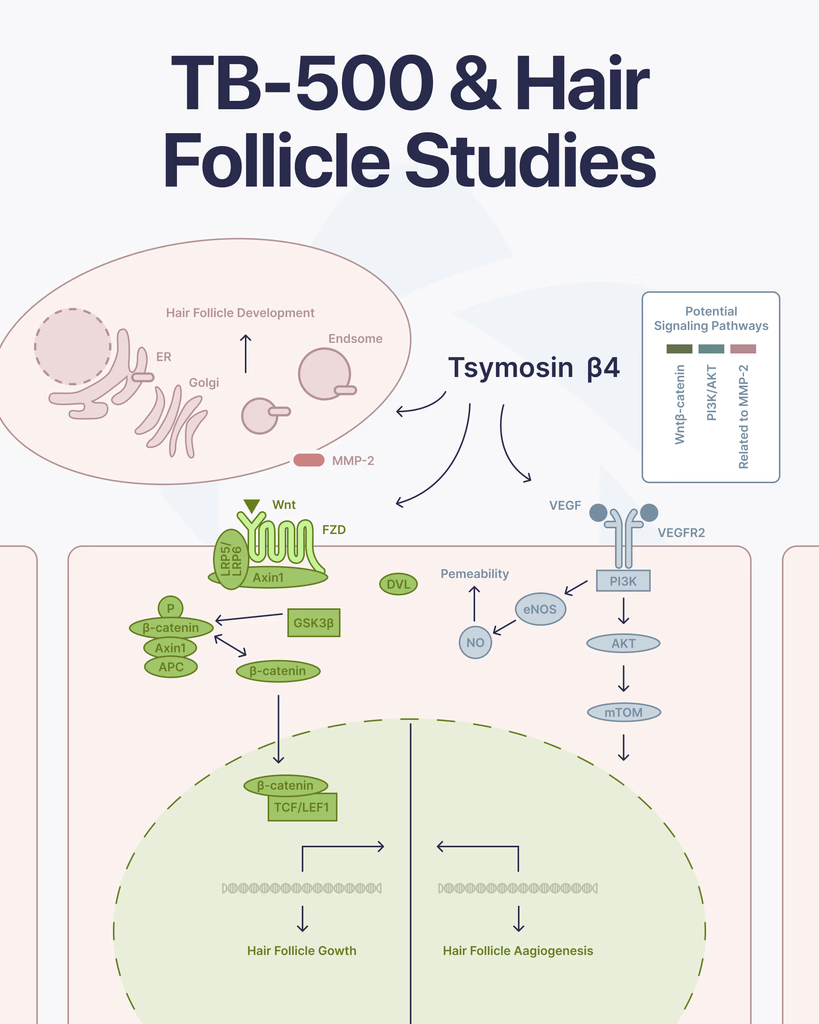
By Dr. Leonard Haberman, Chief Science Officer, OPTMZ Peptides Published: March 2,2026 | Last Updated: April 14, 2026 TB-500 is a synthetic analogue of Thymosin Beta-4 (Tβ4), a 43-amino acid peptide naturally expressed in mammalian tissue. Pre-clinical and in vitro research has examined its role in cell migration, angiogenesis, and tissue repair. More recently, researchers have investigated whether Tβ4-related mechanisms extend to hair follicle biology — an area supported by several published studies, primarily conducted in animal models and isolated cell cultures. What Is TB-500? Molecular Background for Research Contexts TB-500 is derived from the C-terminal fragment of Thymosin Beta-4. Its molecular structure centers on a conserved actin-binding domain (LKKTET sequence), which is understood to regulate actin dynamics — specifically, the polymerization of G-actin into F-actin filaments that govern cellular migration and morphology. In research settings, Tβ4 has been studied across several biological systems, including wound healing models, cardiac tissue, and — relevant to this review — hair follicle regeneration pathways. TB-500’s designation as a synthetic research peptide reflects its use as an accessible laboratory tool for investigating the activity of native Tβ4 in controlled pre-clinical experimental contexts. OPTMZ Peptides supplies TB-500 as a lyophilized research peptide, independently tested by Krause Analytical (DEA-registered, ISO/IEC 17025-certified, Austin TX). Batch purity is verified by HPLC to a minimum of 98%, with most batches returning 98.5–99.9%. Current batch COA data is available in the OPTMZ COA Vault. Hair Follicle Biology: Why Researchers Are Examining Tβ4 Activity Hair follicle cycling involves three principal phases: anagen (active growth), catagen (regression), and telogen (rest). Transition from telogen back into anagen requires coordinated signaling among follicular stem cells, dermal papilla cells, and supporting vascular structures. Disruption at any stage — through reduced stem cell activation, diminished vascularization, or inflammatory signaling — is associated with arrested cycling in research models. Several of the biological pathways Thymosin Beta-4 has been studied for — stem cell migration, vascular endothelial growth factor (VEGF) signaling, and regulation of actin dynamics — overlap with the cellular events that govern this follicular cycle. This mechanistic overlap is the primary reason researchers have examined whether Tβ4 influences hair follicle behavior in experimental contexts. What Studies Have Examined: TB-500 and Hair Follicle Mechanisms Thymosin Beta-4 and Hair Follicle Stem Cell Activation The foundational study in this area was published by Philp et al. (2004) in the FASEB Journal. Researchers investigated the effect of exogenous Thymosin Beta-4 on hair follicle cycling in murine models. The study found that Tβ4 administration was associated with accelerated transition into the anagen phase and attributed this effect, in part, to activation of hair follicle stem cells. The authors concluded that Tβ4 influences critical events in the active phase of the hair follicle cycle, including stem cell mobilization and cell migration toward the dermal papilla (Philp et al., 2004 — PMID 14657002). This study has been cited 166 times and remains the primary reference point for subsequent investigations into Tβ4 and follicular biology. Angiogenesis, VEGF Signaling, and Vascular Support of Follicles Adequate vascularization of the hair follicle bulb is required for nutrient delivery during the anagen phase. Research has examined Thymosin Beta-4’s role in promoting angiogenesis — the formation of new blood vessels from existing vasculature — as a mechanism with potential relevance to follicular support. Philp et al. (2004) in Mechanisms of Development documented that Tβ4 promotes angiogenesis and noted its observed association with hair follicle growth in murine models, connecting the peptide’s vascular activity to follicular biology (Philp et al., 2004 — PMID 15210177). Tβ4’s upregulation of VEGF expression — documented in separate vascular research contexts — is understood to be the mechanistic pathway underlying this angiogenic effect in pre-clinical models. Mechanism Investigation: The 2015 PLOS One Study A 2015 study by Gao et al. published in PLOS One set out specifically to investigate the mechanism by which Thymosin Beta-4 induces hair growth in mouse models. Prior studies had established the association; this study examined the downstream molecular events. The investigators found evidence that Tβ4 influences both stem cell migration and follicular development through pathways involving the Wnt signaling cascade, a known regulator of hair follicle morphogenesis and cycling (Gao et al., 2015 — PMID 26076359). Tβ4 Overexpression and Follicular Morphogenesis Cha et al. (2010) examined what occurs when Thymosin Beta-4 is overexpressed rather than supplemented in controlled models, finding that excess Tβ4 was associated with abnormal hair and tooth morphogenesis — suggesting the peptide operates within regulated concentration ranges relevant to normal follicular architecture (Cha et al., 2010 — PMID 20013654). This finding is methodologically important for researchers designing Tβ4 experimental protocols. Secondary Follicle Research Beyond the primary murine and in vitro models, Dai et al. (2021) examined the role of Thymosin Beta-4 in the biology of secondary hair follicles in goat cashmere production — a distinct but mechanistically relevant model for understanding Tβ4’s influence on follicle initiation and fiber growth pathways (Dai et al., 2021 — PMC7875905). What Does This Research Establish — and What Does It Not? The published pre-clinical literature establishes a coherent mechanistic framework: Thymosin Beta-4 influences hair follicle biology through stem cell activation, angiogenesis, and Wnt pathway modulation in animal models and isolated cell systems. The primary studies are well-cited, methodologically documented, and consistently cited in peer-reviewed reviews of follicular biology. What this research does not establish: Human clinical evidence is limited. The majority of studies have been conducted in murine models or isolated cells. Controlled human trials examining Tβ4 and hair follicle outcomes have not been published at the level of randomized controlled evidence. TB-500 as a synthetic analogue has been studied primarily as a research tool for investigating native Tβ4 activity in pre-clinical settings. Research-grade TB-500 is not a pharmaceutical product, is not FDA-approved, and is supplied exclusively for laboratory use. Dose-response relationships in follicular research remain incompletely characterized. The Cha et al. overexpression data indicates that concentration level is a relevant variable in experimental design. Researchers accessing TB-500 for follicular or related studies should treat the current